Equine Stifle X Ray. To further improve craniocaudal radiographs, get close! stifle radiography in the young performance horse is a fairly routine procedure for most equine practitioners. the stifle is a complex joint and, when faced with a patient suffering from pain in this area, a veterinary surgeon will often use radiography. Department of veterinary clinical sciences, university of melbourne, werribee, victoria 3030,. the stifle joint is an important source of lameness in all types of horses. the equine stifle is a large, complex region and its ultrasonographic examination requires a thorough. With good collimation, the lateromedial view does not require the use of a grid. Radiology of the stifle joint of the horse. radiographs of the stifle in horses are a relatively common procedure. 1,2 because of its clinical importance,. the precise radiographic anatomy of the soft tissue structures of the equine stifle has not been described. The medial tibial condyle should be flat, not curved. If the cartilage or ligaments of the joint have ruptured, a. a radiographic technique is described for the equine stifle joint with the horse in the standing position or under general anaesthesia. a radiographic technique is described for the equine stifle joint with the horse in the standing position or under general.
from veteriankey.com
the stifle joint is an important source of lameness in all types of horses. the aim of this webinar is to discuss the use of different imaging modalities for evaluation of the stifle, describe the relevant osseous and soft tissue anatomy, and to present a practical approach to radiography and ultrasonography of the region. They are used to evaluate for developmental and acquired. The medial tibial condyle should be flat, not curved. a radiographic technique is described for the equine stifle joint with the horse in the standing position or under general. the stifle is a complex joint and, when faced with a patient suffering from pain in this area, a veterinary surgeon will often use radiography. Radiology of the stifle joint of the horse. radiographs of the stifle in horses are a relatively common procedure. a radiographic technique is described for the equine stifle joint with the horse in the standing position or under general anaesthesia. the lateromedial view is the basic radiographic view of the stifle and can be obtained on the standing horse with a portable or fixed radiographic unit.
The Equine Stifle and Tarsus Veterian Key
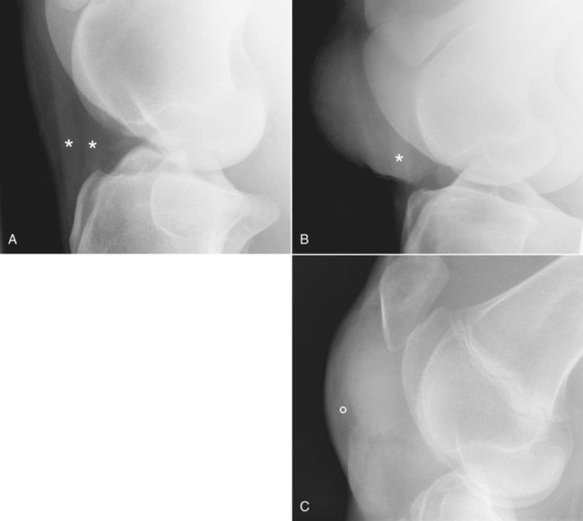
Equine Stifle X Ray the aim of this webinar is to discuss the use of different imaging modalities for evaluation of the stifle, describe the relevant osseous and soft tissue anatomy, and to present a practical approach to radiography and ultrasonography of the region. in severe cases, the horse may carry the leg in a bent position. the stifle joint is an important source of lameness in all types of horses. the ‘take home’ message here is that the equine stifle is prone to problems that can cause significant lameness, and that need. the precise radiographic anatomy of the soft tissue structures of the equine stifle has not been described. the lateromedial view is the basic radiographic view of the stifle and can be obtained on the standing horse with a portable or fixed radiographic unit. the radiographic appearance of the stifle of young horse changes during the first 18 months of life but is not well described in. the aim of this webinar is to discuss the use of different imaging modalities for evaluation of the stifle, describe the relevant osseous and soft tissue anatomy, and to present a practical approach to radiography and ultrasonography of the region. With good collimation, the lateromedial view does not require the use of a grid. Department of veterinary clinical sciences, university of melbourne, werribee, victoria 3030,. The lateral trochlear ridge has lost its regular, rounded appearance and shows an indentation with underlying subchondral bone sclerosis, indicating oc. 1,2 because of its clinical importance,. If the cartilage or ligaments of the joint have ruptured, a. fragments of bone that have pulled away from the attachment sites of the menisci and stifle ligaments can be seen. the stifle is a complex joint and, when faced with a patient suffering from pain in this area, a veterinary surgeon will often use radiography. the equine stifle is a large, complex region and its ultrasonographic examination requires a thorough.